MYOPIA: WHAT IS MYOPIA?
WHAT IS MYOPIA?
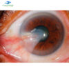
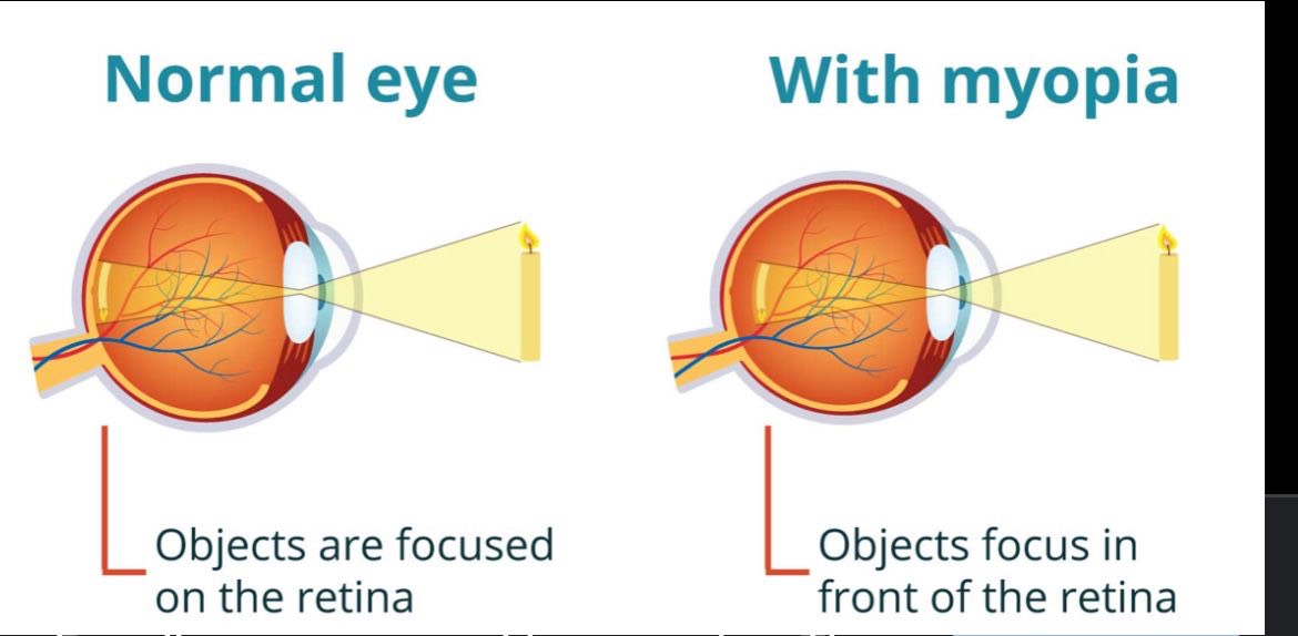
Myopia, also known as near-sightedness or short-sightedness, is an eye disorder where light focuses in front of, instead of focusing on the retina. This causes distant objects to appear blurry while close objects appear normal. Other symptoms may include headaches, eye strain, squinting to see at far, etc. Severe near-sightedness is associated with an increased risk of retinal detachment, cataracts, and glaucoma.
These symptoms may become more obvious when children are between ages 8 and 12 years old.
The underlying mechanism involves the axial length ( length of the eyeball) growing too long or the cornea (the protective outer layer of your eye) is too curved, the light that enters your eye won’t focus correctly. It is a type of refractive error that can be diagnosed by an eye examination.
Tentative evidence indicates that the risk of near-sightedness can be decreased in children by reducing the amount of near work they do, have they spend time outside. This may be related to natural light exposure. Near-sightedness can be corrected with eyeglasses, contact lenses, or refractive surgery. Eyeglasses are the easiest and safest method of correction. Contact lenses can provide a wider field of vision but are associated with a risk of infection. Refractive surgery permanently changes the shape of the cornea.
CAUSES:
Myopia is caused by a cornea that is too steeply curved. It can also be caused by an eye that is longer than normal. In both cases, light rays are focused in front of the retina instead of on it.
The underlying cause is believed to be a combination of genetic and environmental factors.
Risk factors include; doing work that involves focusing on close objects, greater time spent indoors, urbanization, and a family history of the condition. It is also associated with a high socioeconomic class and a higher level of education.
DIAGNOSIS :
A diagnosis of myopia is typically made by an eye care professional, usually an optometrist or ophthalmologist. During refraction, an autorefractor or retinoscope is used to give an initial objective assessment of the refractive status of each eye, then a phoropter is used to subjectively refine the patient’s eyeglass prescription.
MYOPIA PREVALENCE
It is estimated that 34% of the world’s population had myopia in 2020(5.2% with high myopia) and it is predicted that 50% of the world’s population will have myopia by the year 2050(9.8% to have high myopia).
TYPES:
Various forms of myopia have been described by their clinical appearance:
- Simple Myopia: Myopia in an otherwise normal eye, typically less than 4.00 to 6.00 diopters. This is the most common form of myopia.
- Degenerative Myopia: also known as malignant, pathological, or progressive myopia, it is a rare type you usually inherit from your parents. It is characterized by marked fundus changes, such as posterior staphyloma, and associated with a high refractive error and subnormal visual acuity after correction. This form of myopia gets progressively worse over time. Degenerative myopia has been reported as one of the main causes of visual impairment.
- Pseudomyopia: is the blurring of distance vision brought about by a spasm of the accommodation system.
- Nocturnal myopia: Without adequate stimulus for accurate accommodation, the accommodation system partially engages, pushing distance objects out of focus.
- Nearwork-induced transient myopia (NITM): Short-term myopic far point shift immediately following a sustained near visual task.
- Instrument myopia: Over-accommodation when looking into an instrument such as a microscope.
- Induced myopia: also known as acquired myopia, results from various medications, increases in glucose levels, nuclear sclerosis, oxygen toxicity (e.g., from diving or from oxygen and hyperbaric therapy) or other anomalous conditions. Sulphonamide therapy can cause ciliary body edema, resulting in anterior displacement of the lens, pushing the eye out of focus. Elevation of blood-glucose levels can also cause edema (swelling) of the crystalline lens as a result of sorbitol accumulating in the lens. This edema often causes temporary myopia. Scleral buckles, used in the repair of retinal detachments may induce myopia by increasing the axial length of the eye.
- Index myopia is attributed to variation in the index of refraction of one or more of the ocular media. Cataracts may lead to index myopia.
Form deprivation myopia occurs when the eyesight is deprived by limited illumination and vision range, or the eye is modified with artificial lenses or deprived of clear form vision.
DEGREE
The degree of myopia is described in terms of the power of the ideal correction, which is measured in diopters:
- Myopia between-0.00 and -0.50 diopters are usually classified as emmetropia.
- Low myopia usually describes myopia between −0.50 and −3.00 diopters.
- Moderate myopia usually describes myopia between −3.00 and −6.00 diopters.
- High myopia usually describes myopia of −6.00 or more.
Myopia-Related eye problems :
People with myopia are more likely to have retinal detachments and primary open-angle glaucoma. They are also more likely to experience floaters, shadow-like shapes which appear in the field of vision. In addition to this, high myopia is linked to macular degeneration, cataracts, and significant visual impairment.
Age or Onset of Myopia
Myopia is sometimes classified by the age of onset.
- Congenital myopia: also known as infantile myopia, is present at birth and persists through infancy.
- Youth onset myopia occurs in early childhood or teenager, and the ocular power can keep varying until the age of 21.
- School myopia appears during childhood, particularly the school-age years. This form of myopia is attributed to the use of the eyes for close work during the school years.
- Adult-onset myopia
- Early adult-onset myopia occurs between ages 20 and 40.
- Late adult-onset myopia occurs after age 40.
Treatment
Correction using glasses or contact lenses is the most common treatment; other approaches include orthokeratology, and refractive surgery. Medications (mostly atropine) and vision therapy can be effective in addressing the various forms of pseudomyopia.